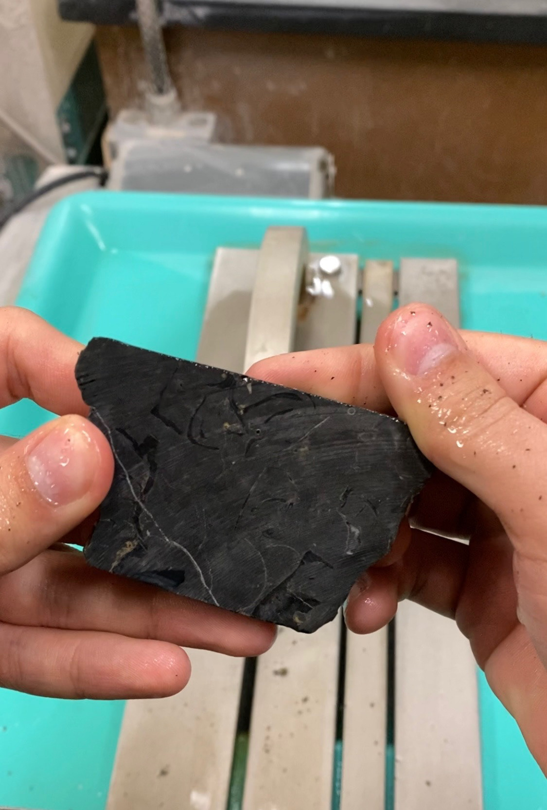
Cut piece of limestone revealing rudist shell fossil within. After being cut, pieces were polished in order to properly image through microscope.
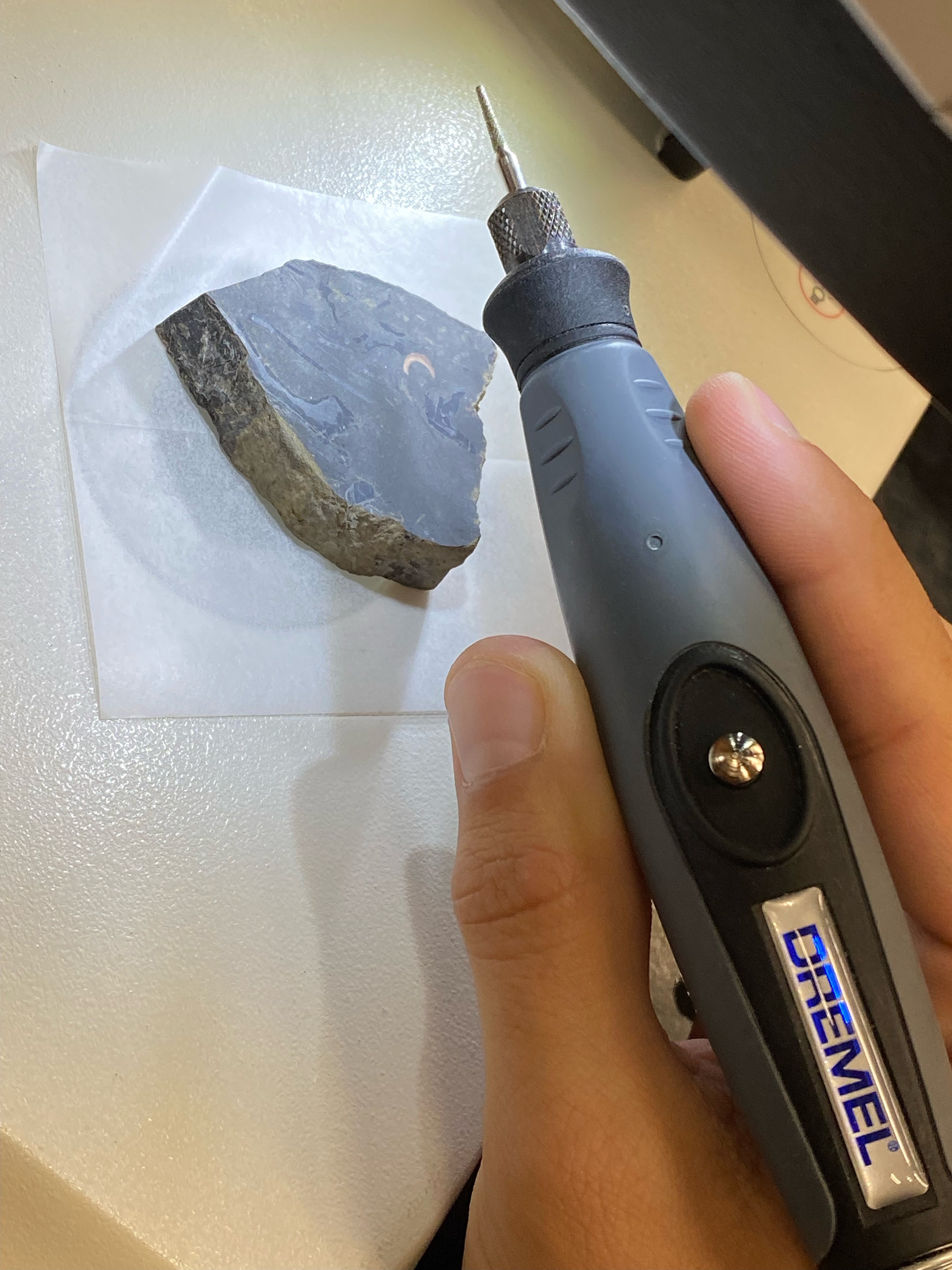
Stage and tool for microsampling. top illumination and an 8x zoom microscope were used to ensure the proper areas were being microsampled.
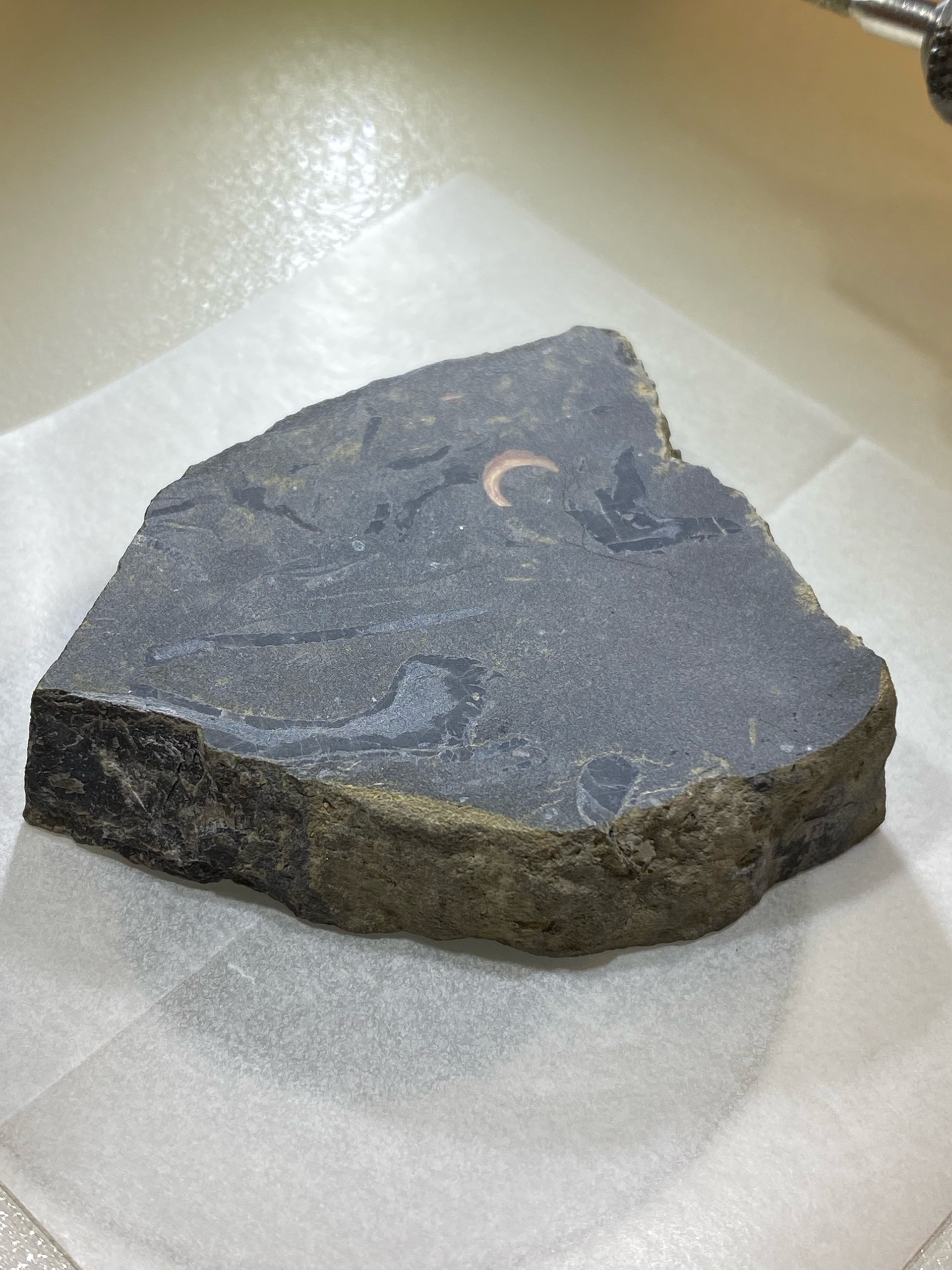
Image of sample just prior to microsampling. The rudist shell is the dark grey on the surface. lighter grey is a matrix/replacement that is untestable. brown object in surface is unknown but also untestable.
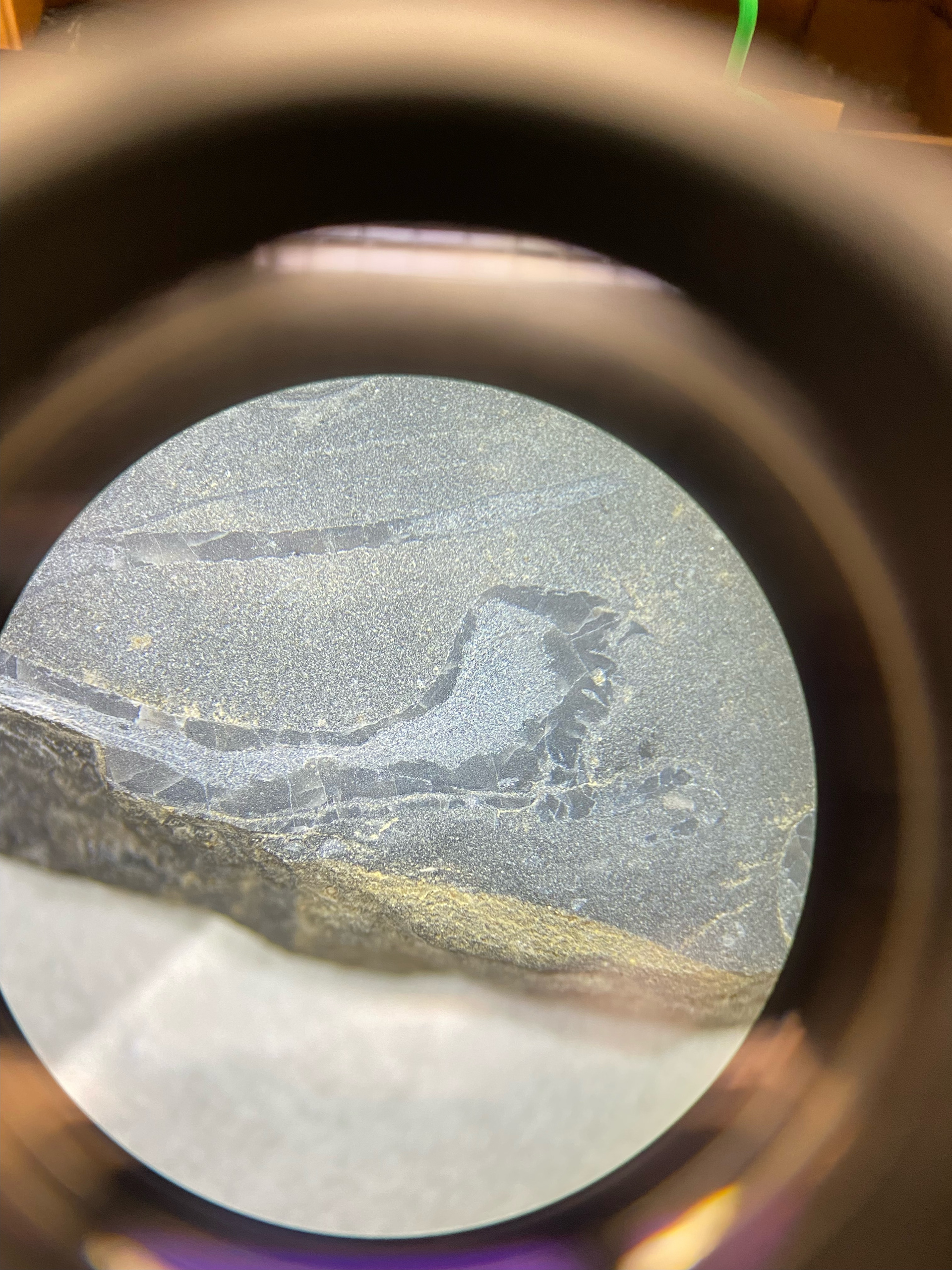
Image of sample through 8x zoom microscope prior to testing. Bivalve sample is the darker grey.
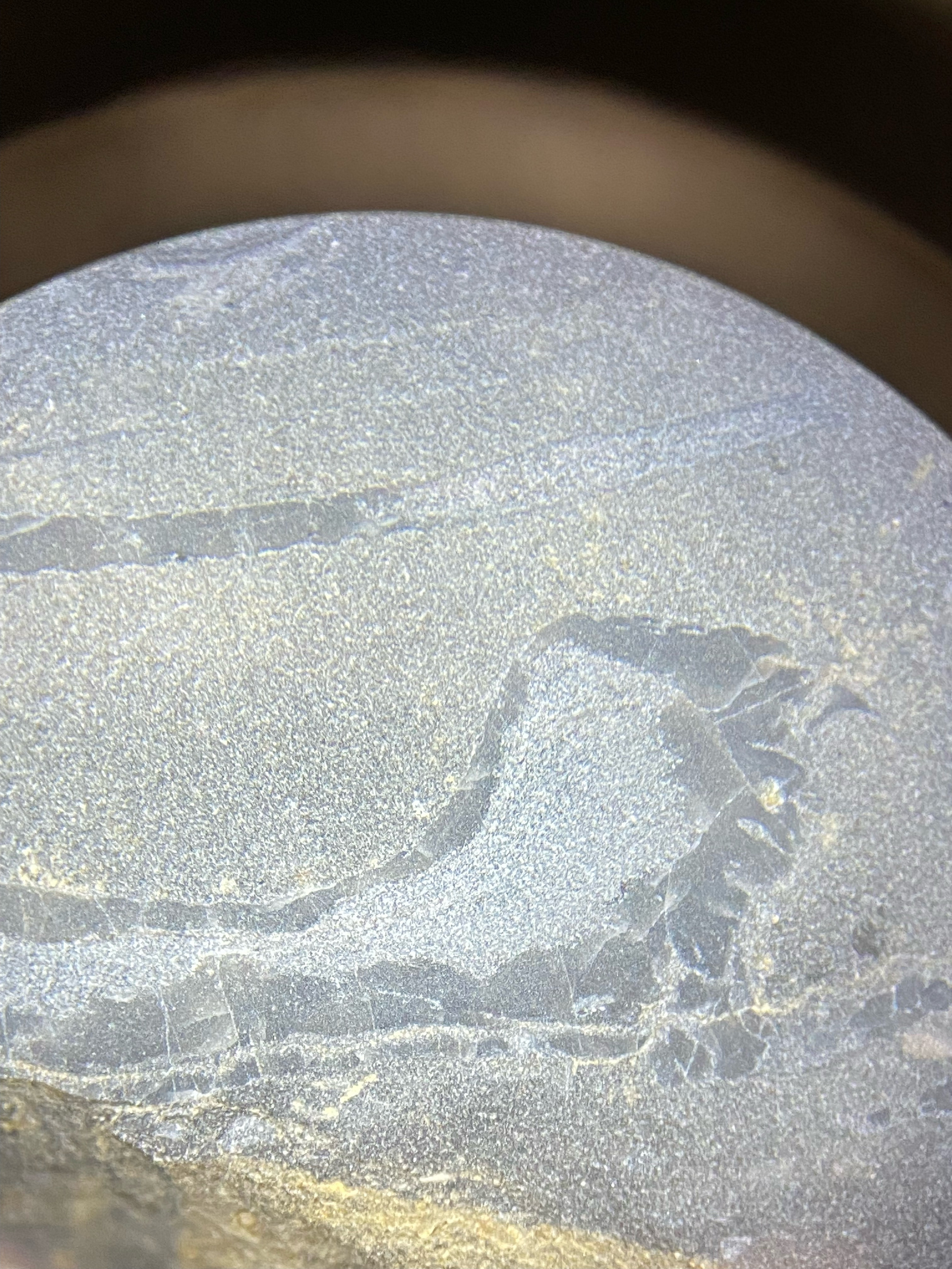
additional photo showing bivalve.
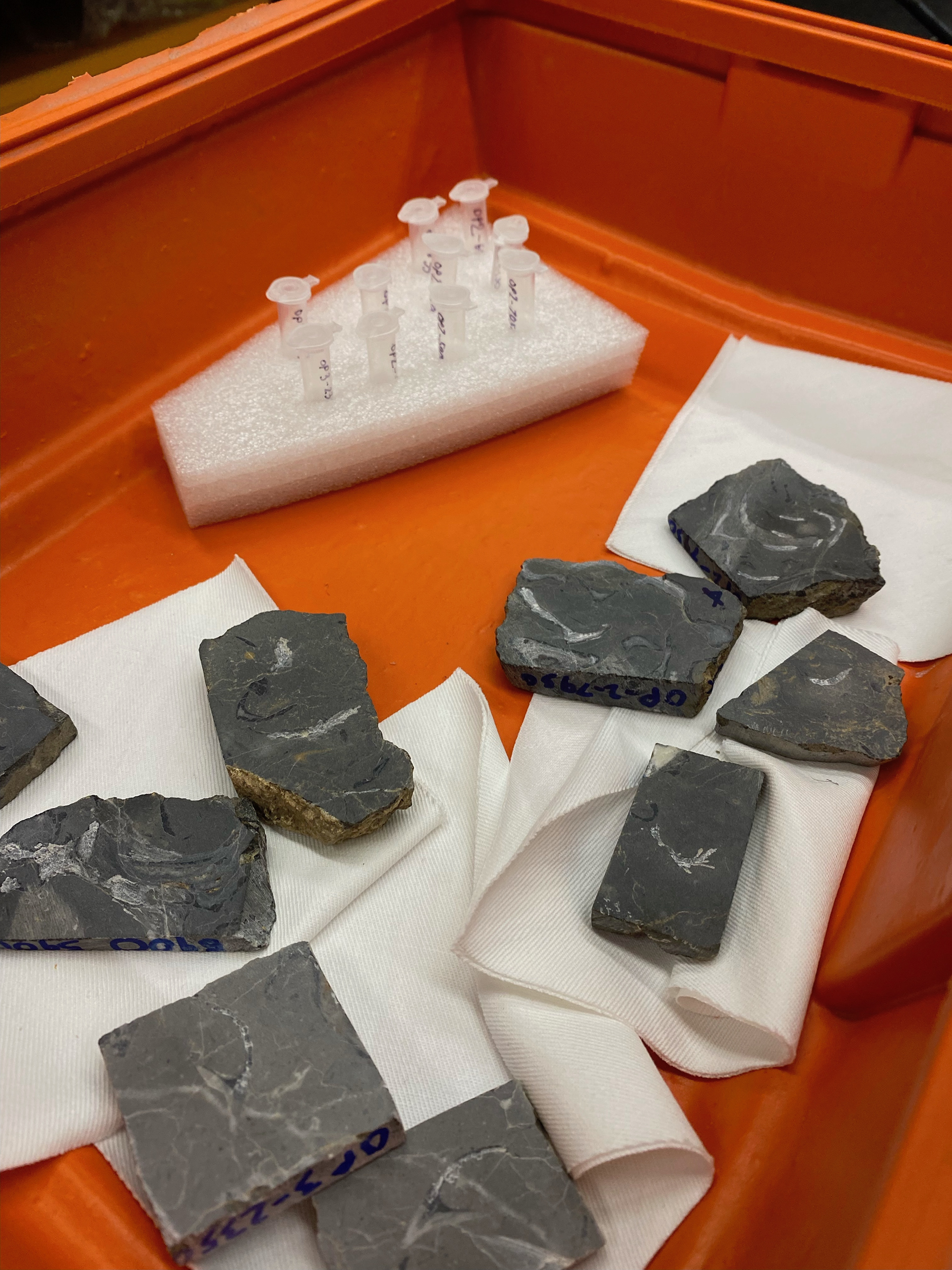
Image of Bivalve fossil rock samples after micro-sampling. Note the scraped out areas within the darker grey that was visible earlier. That region of the sample is the only viable surface of the rock for testing to determine paleoclimate. centrifuge tubes in rear of box are ready to be shipped to the laboratory for testing.